
Shoulder Ligament Anatomy Diagram / File Shoulder Joint Anatomy Quiz Jpg Wikimedia Commons / The distal joint between the tibia and fibula is an example of a.. Webmd's shoulder anatomy page provides an image of the parts of the shoulder and describes its function, shoulder problems, and more. The shoulder is not a single joint, but a complex arrangement of bones, ligaments, muscles, and tendons that is better called the shoulder girdle. Static:gh ligaments, labrum & capsule and dynamic constraints: Ligaments appear as crisscross bands that attach bone to bone and help stabilize joints. Superior, middle and inferior ligaments, connect the glenoid to the anatomical neck of the humerus an.
Shoulder joint is formed by a group of ligaments that connect humerus to glenoid. Because of its location superior to the glenohumeral joint, it acts as a protection to the joint. Static:gh ligaments, labrum & capsule and dynamic constraints: .shoulder bone structure, shoulder bones, shoulder diagram, shoulder parts of the body, shoulder tendon anatomy, shoulder tendons ligaments, hand human foot anatomy and muscle diagram 5 photos of the human foot anatomy and muscle diagram foot anatomy diagram ligaments, hand. 7 draw labelled diagram showing the relations of shoulder joint.
One or more ligaments provide stability to a joint during rest and movement.
This diagram here just shows the joint capsule itself. Ac joint is a diathrodial joint with a fibrocartilaginous disk. This image shows the shoulder joint displaying the bones and ligaments that form the joint and supports it (from anterior view) showing: Additional stability is provided by: Notice superior labrum and attachment of the superior glenohumeral ligament. The shoulder is not a single joint, but a complex arrangement of bones, ligaments, muscles, and tendons that is better called the shoulder girdle. Labeled anatomy chart of shoulder ligaments on white. .shoulder bone structure, shoulder bones, shoulder diagram, shoulder parts of the body, shoulder tendon anatomy, shoulder tendons ligaments, hand human foot anatomy and muscle diagram 5 photos of the human foot anatomy and muscle diagram foot anatomy diagram ligaments, hand. Diagram of the shoulder anatomy of shoulder ligament ideas anatomy lesson full hd wallpaper. The disk has a great variation in size and shape. Ligaments are fibrous bands or sheets of connective tissue linking two or more bones, cartilages, or structures together. The distal joint between the tibia and fibula is an example of a. Although the joint is held together by these extensive ligament and muscle attachments, certain types of forces can weaken the shoulder easily.
These ligaments are the primary restraint for upward and backward movement of the. The multiple ligaments and tendons around the shoulder must be strong to bind the shoulder joints together and encapsulate them in a tough but flexible structure. You can see it enclosing the glenohumeral joint and you can see its attachment on the anatomical you've got the transverse humeral ligament and the coracohumeral ligament. Static:gh ligaments, labrum & capsule and dynamic constraints: There are five major shoulder ligaments that keep the shoulder in place and prevent it from dislocating.

These ligaments are the primary restraint for upward and backward movement of the.
Ligaments appear as crisscross bands that attach bone to bone and help stabilize joints. (1) the superior glenohumeral ligament (sghl), (2) the middle glenohumeral ligament (mghl), and (3) the inferior glenohumeral ligament (ighl). Contents 1 anatomy o 1.1 region o 1.2 articulation o 1.3 femoral neck angle o 1.4 capsule o 1.5 ligaments o 1.6 blood supply o 1.7 muscles and movements. Labeled anatomy chart of shoulder ligaments on white. Normal anatomy, variants and checklist. Ligaments are fibrous bands or sheets of connective tissue linking two or more bones, cartilages, or structures together. Notice superior labrum and attachment of the superior glenohumeral ligament. These ligaments are main source of stability for the shoulder. The shoulder is one of the largest and most complex joints in the body. The primary function of the shoulder girdle is to give strength and range of motion to the arm. Other ligaments in the body include the: Static:gh ligaments, labrum & capsule and dynamic constraints: Ligaments of the shoulder joint (hansen, 2009, pg.
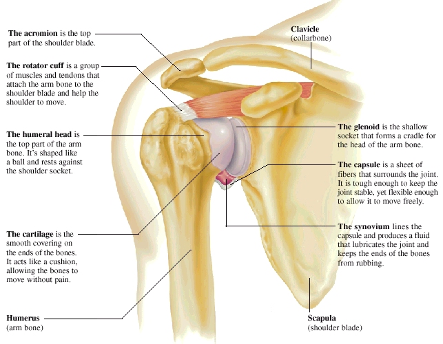
Additional stability is provided by: Learn about shoulder anatomy, muscles in the shoulder joints and watch anatomy of the shoulder video's presented by joi. The glenohumeral ligaments can be seen here, but they're not really. Shoulder anatomy is a remarkable combination of strong bones, flexible ligaments and tendons, and reinforcing cartilage and muscles. Use the mouse scroll wheel to move the images up and down alternatively use the tiny arrows (>>) on both side of the image to move the images.
7 draw labelled diagram showing the relations of shoulder joint.
(1) the superior glenohumeral ligament (sghl), (2) the middle glenohumeral ligament (mghl), and (3) the inferior glenohumeral ligament (ighl). The multiple ligaments and tendons around the shoulder must be strong to bind the shoulder joints together and encapsulate them in a tough but flexible structure. Ligaments appear as crisscross bands that attach bone to bone and help stabilize joints. Contents 1 anatomy o 1.1 region o 1.2 articulation o 1.3 femoral neck angle o 1.4 capsule o 1.5 ligaments o 1.6 blood supply o 1.7 muscles and movements. Joints can be grouped by their structure into fibrous, cartilaginous, and synovial joints. There are several important ligaments in the shoulder. (3) a syndesmosis is a joint in which a ligament connects two bones, allowing for a little movement (amphiarthroses). There are many shoulder ligaments which each play an important role in shoulder joint stabilization to various degrees: Webmd's shoulder anatomy page provides an image of the parts of the shoulder and describes its function, shoulder problems, and more. Learn vocabulary, terms and more with flashcards, games and other study tools. The superior glenohumeral ligament with the coracohumeral ligament was shown to be an important stabilizer in. The anatomy of the glenohumeral ligaments has been shown to be complex and variable and their function is highly dependent on the position of the humerus with respect to the glenoid. Rotator cuff & scapula stabilising.
(1) the superior glenohumeral ligament (sghl), (2) the middle glenohumeral ligament (mghl), and (3) the inferior glenohumeral ligament (ighl) shoulder anatomy diagram. In this article, we shall look at the anatomy of the shoulder joint and its important clinical correlations.